Importance of the renal ion channel TRPM6 in the circadian secretion of renin to raise blood pressure (from Miki-Lab, in Nat. Commun.)
Blood pressure has a daily pattern, with higher values in the active period. Its elevation at the onset of the active period substantially increases the risk of fatal cardiovascular events. Renin secretion stimulated by renal sympathetic neurons is considered essential to this process; however, its regulatory mechanism remains largely unknown. Here, we show the importance of transient receptor potential melastatin-related 6 (TRPM6), a Mg2+-permeable cation channel, in augmenting renin secretion in the active period. TRPM6 expression is significantly reduced in the distal convoluted tubule of hypotensive Cnnm2-deficient mice. We generate kidney-specific Trpm6-deficient mice and observe a decrease in blood pressure and a disappearance of its circadian variation. Consistently, renin secretion is not augmented in the active period. Furthermore, renin secretion after pharmacological activation of β-adrenoreceptor, the target of neuronal stimulation, is abrogated, and the receptor expression is decreased in renin-secreting cells. These results indicate crucial roles of TRPM6 in the circadian regulation of blood pressure.
This article was published in Nature Communications on June 17, 2021.
Title: “Importance of the renal ion channel TRPM6 in the circadian secretion of renin to raise blood pressure”
Authors: Yosuke Funato, Daisuke Yamazaki, Daisuke Okuzaki, Nobuhiko Yamamoto, and Hiroaki Miki
Links
-
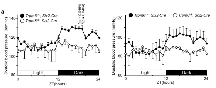
Blood pressure values of 2- to 3-month-old mice of the indicated genotypes were measured with radiotelemetry. Data are shown as means ± SEM for systolic (left) and diastolic (right) pressure values (n = 4 for Trpm6+/+; Six2-Cre and n = 3 for Trpm6fl/fl; Six2-Cre). The p-values were determined by two-way ANOVA with Holm-Sidak post hoc tests. *p < 0.05.
-
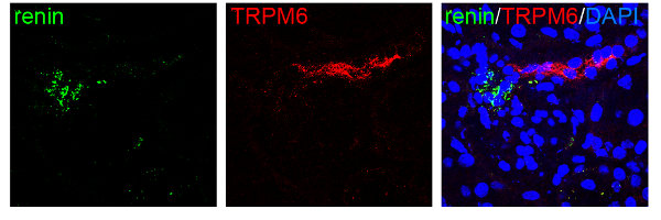
Cryosections of 2-month-old Trpm6+/+; Six2-Cre mouse kidneys were subjected to immunofluorescence staining with the indicated antibodies and DAPI (blue). Merged images and monochrome images of each antibody staining are shown.
- Home
- Achievement
- Research Activities
- Importance of the renal ion channel TRPM6 in the circadian secretion of renin to raise blood pressure (from Miki-Lab, in Nat. Commun.)