Uncovering Critical Flaws in the Detection and Elimination of Senescent Cells Using the p16-3MR Mouse Model (Hara Lab, in EMBO Reports)
A research group led by Professor Eiji Hara and Graduate student Nozomi Hori (at the time) at the Research Institute for Microbial Diseases, The University of Osaka, together with Associate Professor Shimpei Kawamoto (currently Professor at the Center for Healthspan Research, Tohoku University), has identified functional deficiencies in the p16-3MR mouse model, which has been widely used in cellular senescence research.
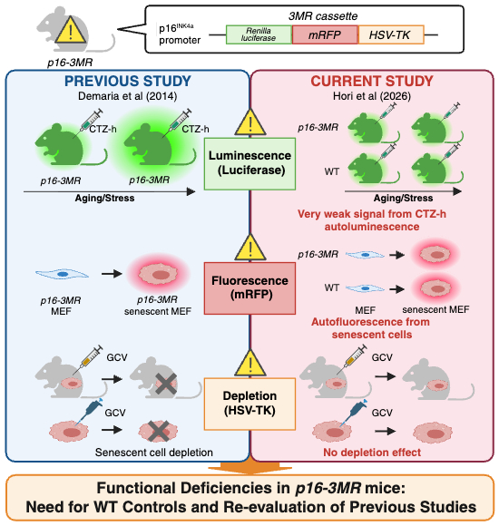
Cellular senescence plays an important role as a tumor suppression mechanism; however, senescent cells accumulate in tissues with advancing age and contribute to the development of various aging-related diseases through the Senescence-Associated Secretory Phenotype (SASP), a program involving the secretion of numerous pro-inflammatory factors. Mouse models that enable the in vivo visualization and selective elimination of senescent cells are therefore indispensable tools for elucidating the functions of senescent cells in living organisms. The p16-3MR mouse was designed to express a trimodality reporter fusion protein (3MR) - comprising functional domains of Renilla luciferase (Rluc), monomeric red fluorescent protein (mRFP), and herpes simplex virus 1 thymidine kinase (HSV-TK) - under the p16INK4a gene promoter. Since its first description by Demaria et al. (Developmental Cell, 2014), this model has been one of the most widely used mouse models in senescence research, enabling both visualization and selective elimination of senescent cells through ganciclovir (GCV) administration.
In the present study, the authors demonstrated that bioluminescence imaging (BLI) signals in p16-3MR mice are extremely weak and virtually indistinguishable from the background signals observed in wild-type (WT) mice injected with coelenterazine-h (CTZ-h), the substrate for Rluc. Under conditions of aging, doxorubicin treatment, and cutaneous wound healing, no significant changes in BLI signals were detected, and the results reported by Demaria et al. (2014) could not be reproduced. Furthermore, mRFP signals were undetectable in senescent fibroblasts derived from p16-3MR mice, and GCV treatment failed to eliminate senescent cells. In addition, GCV was shown to affect immune cell populations such as macrophages in an HSV-TK-independent manner, highlighting the danger of interpreting the effects of GCV treatment as selective elimination of senescent cells in the absence of WT controls.
A key strength of this study is the systematic verification of functional deficiencies in the 3MR transgene through multiple independent approaches, including side-by-side comparative experiments using p16-3MR mice obtained from both the Campisi and Demaria laboratories, whole-genome sequencing to verify the transgene sequence, and experiments using albino p16-3MR mice. Collectively, these results demonstrate functional deficiencies in all three components of the 3MR transgene and underscore the importance of reproducibility and rigorous experimental design in senescence research.
While the results of this study do not definitively conclude that the p16-3MR mouse model is completely non-functional, they demonstrate that WT mice must always be included as appropriate negative controls when using this model, and suggest that caution is warranted in interpreting the findings of previously published studies that employed p16-3MR mice. It is hoped that this study will serve as a foundation for establishing more reliable methodological standards in senescent cell research.
It should be noted that Professor Eiji Hara and Professor Naoko Ohtani (Graduate School of Medicine, Osaka Metropolitan University) are co-authors of Demaria et al. (Developmental Cell, 2014), the study examined in this work. However, neither author conducted any experiments using the p16-3MR mouse model in that study, and all remaining authors declare no conflicts of interest.
This study was published online in EMBO Reports on May 28, 2026.
Title: Limitations of the p16-3MR mouse model for detecting and eliminating senescent cells
Authors: Nozomi Hori, Shimpei Kawamoto, Ken Uemura, Yumi Kinugasa-Katayama, Yumiko Okumura, Kentaro Tanaka, Jeong Hoon Park, Masahiro Wakita, Daisuke Motooka, Naoko Ohtani & Eiji Hara
DOI: 10.1038/s44319-026-00802-8
Links
- Home
- Achievement
- Research Activities
- Uncovering Critical Flaws in the Detection and Elimination of Senescent Cells Using the p16-3MR Mouse Model (Hara Lab, in EMBO Reports)