Ikawa Lab/Bioinformatics Center Department of Experimental Genome Research
In the past, animals harboring natural mutations have been used to elucidate the mechanisms underlying various diseases.
In the “post-genome project era,” genetically modified animals play a key role in basic molecular biological investigations and act as models of human disease. Our laboratory studies the mechanisms underlying mammalian reproductive systems through genetic manipulation of animal models.
Analysis of molecular mechanisms involved in mammalian reproduction
Our laboratory focuses on mechanistically studying the mammalian reproduction system in vivo using gene-manipulated animals. We were the first laboratory in the world to produce genetically modified mice that express a green fluorescent protein (GFP) throughout the body (Figure 1, FEBS Lett 1997). These green fluorescent mice are useful for many types of research projects. Indeed, we used these animals to label sperm with a fluorescent protein and visualized the fertilization process (Figure 1, Exp Anim 2010)
We introduced the cutting-edge CRISPR/Cas9 system and have been improving the technology (SciRep 2013, 2016; Science 2018). By utilizing the system, we have elucidated the lumicrine system; testis-derived NELL2 goes through the male reproductive tract's luminal space and triggers differentiation of epididymal epithelial cells through ROS1 receptor kinase. Then, activated epididymal cells secrete OVCH2 protease that modulates sperm fertilizing ability by trimming sperm membrane ADAM3 protein (Science 2020). We also found that sperm calcineurin (PPP3CC/PPP3R2) is essential for sperm motility and male fertility (Figure 2, Science 2015). Inhibiting these pathways may lead to the development of a reversible male contraceptive (Andrology 2025).
More recently, besides IZUMO1 (Nature 2005), we found novel sperm proteins essential for the sperm-oocyte fusion process (PNAS 2021, 2022, 2023, Cell 2024). Our laboratory will continue elucidating the mammalian fertilization mechanism.
Development of new technologies for producing genetically modified animals
We are also developing new reproductive research tools and assisted reproductive technologies using various viral vectors and tissue culture devices.
For example, we have successfully developed a mouse model of preeclampsia by genetically modifying only the placenta using a lentiviral vector (Figure 3, Nat Technol 2007). Recently, we have also been conducting research utilizing the advantage of transient gene expression by using adenoviral vectors and LNP (Lipid NanoParticle). Additionally, by combining gas-permeable membranes and porous membranes to culture testes, we have successfully reproduced and observed spermatogenesis in vitro (Sci Rep 2025). We have also reproduced the implantation in vitro (Nat Commun 2025). By combining these techniques, we aim to elucidate mechanisms of reproduction and develop assisted reproductive technologies.
Our laboratory and the Animal Resource Center for Infectious Diseases support services such as the generation of genetically modified animals, in vitro fertilization, and cryopreservation of mouse strains.
For more information about our research and services, please visit our homepage (https://egr.biken.osaka-u.ac.jp/).
-
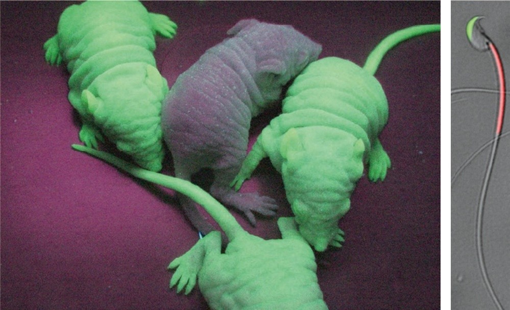
Fig. 1. GFP-expressing mice. Our “Green mice” have been used by hundreds of researchers and are good models for studying human disease (FEBS Lett 1997). Fig. 2. RBGS sperm. Transgenic spermatozoa carrying GFP and dDsRed2 in their acrosome and mitochondria. These gametes are useful to visualize the fertilization process (Exp Anim 2010).
-
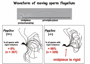
Fig. 2. Calcineurin-deficient sperm. Sperm calcineurin is required for sperm motility for successful fertilization (Science 2015).
-
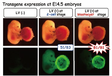
Fig. 3. Lentiviral vector-mediated transgenesis in mice. Lentiviral vectors cannot transduce eggs with zona pellucida (ZP) (left). Without ZP, transductions of fertilized egg and blastocyst result in the whole transgenic (middle) and placenta-specific transgenic (right), respectively (Nat Biotechnol 200).
Staff
- Prof.: Masahito Ikawa
- Assoc. Prof.: Haruhiko Miyata(concur.)
- Asst. Prof.: Chihiro Emori
- Asst. Prof.: Yuki Hatanaka
- Asst. Prof.: Daisuke Mashiko(concur.)
- SA Asst. Prof.: Rie Iida(concur.)
- SA Asst. Prof.: Shingo Tonai(concur.)
- SA Asst. Prof.: Yu Ishikawa
- Postdoc.: Seiya Oura
- Postdoc.: Natsuki Mikami
- Postdoc.: WANG HAOTING
Website
Publications
1. Formation of a complex between TMEM217 and the sodium-proton exchanger SLC9C1 is crucial for mouse sperm motility and male fertility. Iida-Norita et al., PNAS (2025) PMID: 41091759
2. Sperm and offspring production in a nonobstructive azoospermia mouse model via testicular mRNA delivery using lipid nanoparticles. Mashiko et al., PNAS (2025) PMID: 41082659
3. GALNTL5 binds GalNAc and is required for migration through the uterotubal junction and sperm-zona pellucida binding. Noda & Uriu et al., Nat Commun (2025) PMID: 40962834
4. Proximity labeling of axonemal protein CFAP91 identifies EFCAB5 that regulates sperm motility. Wang et al., Nat Commun (2025) PMID: 40931011
5. An ex vivo uterine system captures implantation, embryogenesis, and trophoblast invasion via maternal-embryonic signaling. Hiraoka et al., Nat Commun (2025) PMID: 40595542
6. TEX38 localizes ZDHHC19 to the plasma membrane and regulates sperm head morphogenesis in mice. Kaneda et al., PNAS (2025) PMID: 40030029
7. A conserved fertilization complex bridges sperm and egg in vertebrates. Deneke et al., Cell (2024) PMID: 39423812
8. ZP2 cleavage blocks polyspermy by modulating the architecture of the egg coat. Nishio et al., Cell (2024) PMID: 38490181
9. A small secreted protein NICOL regulates lumicrine-mediated sperm maturation and male fertility. Kiyozumi et al., Nat. Commun. (2023) PMID: 37095084
10. TSKS localizes to nuage in spermatids and regulates cytoplasmic elimination during spermiation. Shimada et al., PNAS (2023) PMID: 36881620
11. 1700029I15Rik orchestrates the biosynthesis of acrosomal membrane proteins required for sperm–egg interaction. Lu et al., PNAS (2023) PMID: 36787362
12. Testis-enriched ferlin, FER1L5, is required for Ca2+-activated acrosome reaction and male fertility. Morohoshi et al., Science Adv. (2023) PMID: 36696506
- Home
- Laboratories
- Ikawa Lab